The relationship between high blood pressure and leg swelling represents a complex interplay of cardiovascular, renal, and vascular mechanisms that affects millions of people worldwide. While hypertension doesn’t directly cause peripheral oedema in most cases, the physiological changes associated with elevated blood pressure can contribute to fluid retention and swelling in the lower extremities. Understanding this connection becomes particularly crucial when you consider that approximately 1.13 billion people globally suffer from hypertension, and many experience concurrent lower limb swelling that may be attributed to various underlying mechanisms.
The distinction between direct causation and secondary effects is essential for proper diagnosis and treatment. Hypertension-related leg swelling often results from the medications used to treat high blood pressure, particularly calcium channel blockers, rather than the elevated blood pressure itself. However, when hypertension leads to complications such as heart failure or kidney disease, these conditions can indeed manifest as significant peripheral oedema, creating a cascade of cardiovascular consequences that require immediate medical attention.
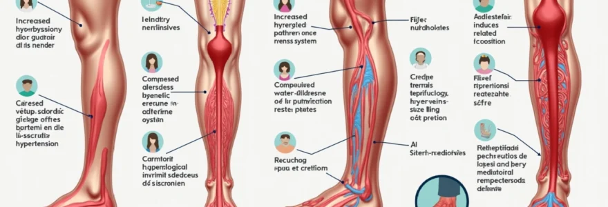
Hypertension-induced peripheral oedema: pathophysiological mechanisms
The development of leg swelling in hypertensive patients involves multiple interconnected pathophysiological pathways that extend far beyond simple fluid retention. Chronic hypertension creates a sustained state of increased vascular pressure that affects the delicate balance between fluid filtration and reabsorption at the capillary level. This disruption occurs through Starling forces, where elevated hydrostatic pressure overwhelms the oncotic pressure gradient, leading to increased fluid extravasation into the interstitial tissues of the lower extremities.
The renin-angiotensin-aldosterone system (RAAS) plays a pivotal role in this process, as chronic hypertension often involves dysregulation of this hormonal pathway. When blood pressure remains consistently elevated, the kidneys may respond by retaining sodium and water, attempting to maintain adequate perfusion pressure. This compensatory mechanism, while initially protective, ultimately contributes to volume overload and subsequent peripheral oedema formation, particularly in gravity-dependent areas such as the ankles and feet.
Increased hydrostatic pressure in lower extremity venous system
Elevated systemic blood pressure directly translates to increased venous pressure throughout the circulatory system, with the lower extremities bearing the brunt of gravitational effects. This increased hydrostatic pressure in the venous system creates a pressure gradient that favours fluid movement from the intravascular space into the surrounding tissues. The phenomenon becomes particularly pronounced during prolonged standing or sitting, when venous return mechanisms are compromised.
The venous valves, which normally prevent retrograde blood flow, may become incompetent under sustained pressure, leading to venous stasis and further fluid accumulation. This process is exacerbated by the fact that hypertensive patients often develop structural changes in their blood vessels , including increased wall thickness and reduced compliance, which impairs the natural pumping action of the venous system.
Compromised lymphatic drainage due to elevated systemic pressure
The lymphatic system, responsible for draining excess interstitial fluid, can become overwhelmed when faced with increased fluid production secondary to hypertension. Elevated systemic pressure affects lymphatic vessels similarly to blood vessels, potentially reducing their drainage capacity and contributing to fluid accumulation in the tissues. Research indicates that chronic hypertension may lead to structural changes in lymphatic vessels, including reduced contractility and impaired valve function.
This lymphatic dysfunction becomes particularly problematic in the lower extremities, where lymphatic drainage must work against gravity. When combined with increased capillary filtration due to elevated blood pressure, the result is a net positive fluid balance in the interstitial space, manifesting as visible swelling in the feet, ankles, and lower legs.
Sodium and water retention through Renin-Angiotensin-Aldosterone system activation
The RAAS activation in hypertensive patients creates a cascade of hormonal changes that promote sodium and water retention. Angiotensin II, a potent vasoconstrictor produced in this system, not only increases blood pressure but also stimulates aldosterone release from the adrenal cortex. Aldosterone, in turn, promotes sodium reabsorption in the kidneys, leading to increased plasma volume and subsequently elevated hydrostatic pressure throughout the vascular system.
This mechanism becomes self-perpetuating, as increased plasma volume contributes to higher blood pressure, which further activates the RAAS. The resulting fluid overload manifests primarily in the dependent portions of the body, creating the characteristic bilateral leg swelling seen in many hypertensive patients. The severity of oedema often correlates with the degree of RAAS activation and the duration of untreated hypertension.
Capillary permeability changes in hypertensive patients
Chronic hypertension induces structural and functional changes in the microvasculature that can increase capillary permeability. These changes include endothelial dysfunction, increased oxidative stress, and inflammation, all of which compromise the normal barrier function of capillary walls. When capillary permeability increases, protein-rich fluid leaks more readily into the interstitial space, reducing the oncotic pressure gradient that normally keeps fluid within the vascular compartment.
The increased permeability particularly affects the lower extremities due to the combined effects of gravity and higher hydrostatic pressure in dependent areas. This pathophysiological change explains why some hypertensive patients develop oedema even when their cardiac function remains normal and their medication regimen doesn’t include agents typically associated with fluid retention.
Clinical manifestations of hypertensive leg swelling vs other aetiologies
Distinguishing hypertension-related leg swelling from other causes requires careful clinical assessment and understanding of the characteristic features associated with different aetiologies. The presentation of oedema can provide valuable diagnostic clues, helping healthcare providers identify the underlying pathophysiological mechanisms and guide appropriate treatment strategies. Hypertensive leg swelling typically presents with specific patterns and characteristics that differentiate it from cardiac, renal, or venous causes of peripheral oedema.
The temporal pattern of swelling onset and progression often differs significantly between hypertensive and non-hypertensive causes. While acute heart failure may present with rapid-onset severe oedema accompanied by dyspnoea, hypertension-related swelling typically develops gradually over weeks to months. The distribution of oedema also varies, with hypertensive swelling usually affecting both legs symmetrically, starting from the ankles and progressing upward with increasing severity of the underlying condition.
Bilateral pitting oedema characteristics in essential hypertension
Essential hypertension-related oedema typically manifests as bilateral, symmetrical pitting oedema that begins in the most dependent parts of the body. The swelling characteristically worsens throughout the day due to gravitational effects and improves with leg elevation overnight. When you press firmly on the swollen area for several seconds, it leaves a temporary indentation or “pit,” hence the term pitting oedema. This characteristic distinguishes it from non-pitting oedema seen in conditions such as lymphoedema or myxoedema.
The severity of pitting oedema in hypertensive patients often correlates with the degree of blood pressure elevation and the presence of target organ damage. Patients with well-controlled hypertension rarely develop significant oedema unless they’re taking medications that promote fluid retention. The oedema typically responds well to appropriate antihypertensive therapy and lifestyle modifications , including dietary sodium restriction and regular physical activity.
Distinguishing heart Failure-Related oedema from hypertensive swelling
Heart failure represents a common complication of long-standing hypertension, and distinguishing between simple hypertensive oedema and heart failure-related fluid retention is crucial for appropriate management. Heart failure-related oedema typically presents with additional symptoms such as dyspnoea on exertion, orthopnoea, paroxysmal nocturnal dyspnoea, and fatigue. The swelling in heart failure patients is often more severe and may progress to involve the thighs, abdomen, and even the upper extremities in advanced cases.
Jugular venous distension, hepatomegaly, and third heart sounds (S3 gallop) are clinical findings that suggest heart failure rather than simple hypertensive oedema. Additionally, chest X-rays in heart failure patients may show cardiomegaly, pulmonary vascular redistribution, or frank pulmonary oedema, findings that are absent in patients with isolated hypertensive leg swelling. Brain natriuretic peptide (BNP) or N-terminal pro-BNP levels can provide valuable diagnostic information, as they are significantly elevated in heart failure but remain normal in patients with simple hypertensive oedema.
Chronic venous insufficiency differential diagnosis
Chronic venous insufficiency (CVI) can coexist with hypertension, making the differential diagnosis challenging in some patients. However, CVI typically presents with specific clinical features that help distinguish it from purely hypertensive oedema. These include skin changes such as hyperpigmentation, varicose veins, venous ulcers, and a characteristic distribution of swelling that may be unilateral or more prominent on one side.
The oedema associated with CVI often has a “woody” or indurated quality, particularly in chronic cases, and may be accompanied by skin thickening and subcutaneous fibrosis. Unlike hypertensive oedema, which typically improves significantly with leg elevation, CVI-related swelling may only partially resolve with elevation and often requires compression therapy for optimal management. Doppler ultrasound examination can definitively diagnose venous insufficiency by demonstrating reflux in the affected venous segments.
Medication-induced oedema from calcium channel blockers
Calcium channel blockers, particularly dihydropyridine agents such as amlodipine and nifedipine, are among the most common causes of medication-induced leg swelling in hypertensive patients. The mechanism involves preferential arterial vasodilation without corresponding venodilation, creating increased capillary pressure and subsequent fluid extravasation. This type of oedema typically develops within weeks to months of starting the medication and affects up to 10-15% of patients taking these agents.
The swelling caused by calcium channel blockers is characteristically bilateral, non-pitting initially, and predominantly affects the ankles and feet. Unlike heart failure-related oedema, it’s not associated with dyspnoea or other cardiac symptoms. The oedema typically resolves within days to weeks after discontinuing the offending medication, though switching to a non-dihydropyridine calcium channel blocker or adding an ACE inhibitor may allow continued use of the medication while reducing oedema severity.
Antihypertensive medications associated with lower limb oedema
The pharmacological management of hypertension involves various drug classes, each with distinct mechanisms of action and potential side effects. Understanding which antihypertensive medications can cause leg swelling is essential for both healthcare providers and patients, as medication-induced oedema represents one of the most common causes of lower limb swelling in the hypertensive population. The incidence and severity of medication-induced oedema vary significantly between different drug classes , with some agents having a much higher propensity to cause fluid retention than others.
Calcium channel blockers, particularly the dihydropyridine subclass, are the most frequent culprits of antihypertensive medication-induced leg swelling. These medications work by blocking calcium channels in vascular smooth muscle cells, leading to vasodilation and reduced peripheral resistance. However, their preferential effect on arterial vessels without corresponding venous dilation creates an imbalance in pre-capillary and post-capillary resistance, resulting in increased capillary hydrostatic pressure and subsequent oedema formation.
Studies indicate that dihydropyridine calcium channel blockers cause peripheral oedema in approximately 2-15% of patients, with the incidence being dose-dependent and more common in elderly patients and women.
Beta-blockers, while generally well-tolerated, can occasionally cause fluid retention, particularly in patients with underlying heart failure or compromised cardiac function. Non-selective beta-blockers may have a higher propensity to cause oedema compared to cardioselective agents, as they can affect peripheral vascular tone and potentially compromise venous return mechanisms. The mechanism involves reduced cardiac output and potential peripheral vasoconstriction, which may contribute to fluid retention in susceptible individuals.
Alpha-blockers such as doxazosin and prazosin can cause oedema through their vasodilatory effects, though this side effect is less common compared to calcium channel blockers. The mechanism involves peripheral vasodilation that can lead to fluid extravasation, particularly when combined with other factors such as prolonged standing or concurrent use of other vasodilating medications. The risk of oedema with alpha-blockers is often related to the degree of blood pressure reduction and individual patient susceptibility factors.
Direct vasodilators like hydralazine and minoxidil are potent causes of fluid retention and peripheral oedema, often requiring concurrent use of diuretics to manage this side effect effectively. These medications cause profound arterial vasodilation, leading to compensatory activation of the renin-angiotensin-aldosterone system and subsequent sodium and water retention. The oedema associated with these agents can be quite severe and may limit their clinical use in some patients.
Diagnostic assessment methods for Hypertension-Related leg swelling
The diagnostic evaluation of leg swelling in hypertensive patients requires a systematic approach that considers both cardiovascular and non-cardiovascular causes of oedema. The assessment begins with a comprehensive history and physical examination, focusing on the temporal pattern of swelling, associated symptoms, medication history, and signs of underlying cardiac, renal, or hepatic dysfunction. A thorough medication review is particularly crucial , as many antihypertensive agents can contribute to peripheral oedema through various mechanisms.
Physical examination should include assessment of the degree and distribution of oedema, presence of jugular venous distension, cardiac auscultation for murmurs or gallops, pulmonary examination for signs of congestion, and abdominal examination for hepatomegaly or ascites. The presence of unilateral swelling, skin changes, or varicose veins may suggest venous insufficiency rather than systemic causes of oedema. Pitting versus non-pitting oedema characteristics can provide valuable diagnostic clues about the underlying aetiology.
Laboratory investigations form an essential component of the diagnostic workup and should include a complete blood count, comprehensive metabolic panel, liver function tests, thyroid function studies, and urinalysis with microscopy. Brain natriuretic peptide (BNP) or N-terminal pro-BNP measurement can help distinguish cardiac from non-cardiac causes of oedema, with elevated levels strongly suggesting heart failure. Serum albumin levels are important to assess for hypoproteinaemia, which can contribute to oedema formation through reduced oncotic pressure.
Imaging studies may be necessary depending on the clinical presentation and initial test results. Echocardiography is indicated when heart failure is suspected, providing information about cardiac structure and function, including ejection fraction, wall motion abnormalities, and valvular function. Chest X-rays can reveal cardiomegaly, pulmonary vascular redistribution, or pleural effusions. Doppler ultrasound of the lower extremities should be considered when venous insufficiency is suspected, as it can identify venous reflux and rule out deep vein thrombosis.
Recent guidelines emphasise the importance of BNP or NT-proBNP testing in patients presenting with dyspnoea and oedema, as these biomarkers have high negative predictive value for excluding heart failure when levels are below established thresholds.
Advanced imaging techniques such as cardiac magnetic resonance imaging (MRI) may be considered in cases where echocardiography provides inconclusive results or when infiltrative cardiomyopathy is suspected. Renal imaging with ultrasound or CT may be appropriate when chronic kidney disease is suspected as a contributing factor. The diagnostic approach should be tailored to individual patient characteristics, with consideration for age, comorbidities, and severity of symptoms.
Secondary hypertension causes contributing to peripheral oedema
Secondary hypertension, representing approximately 5-10% of all hypertension cases, encompasses various underlying conditions that can directly contribute to both elevated blood pressure and peripheral oedema. These conditions often have specific pathophysiological mechanisms that promote fluid retention, making the relationship between hypertension and leg swelling more direct than in essential hypertension. Identifying secondary causes is crucial because treating the underlying condition often leads to resolution of both hypertension and associated oedema .
Primary aldosteronism (hyperaldosteronism) represents one of the most common forms of secondary hypertension, affecting 5-13% of hypertensive patients. This condition involves excessive production of aldosterone by the adrenal glands, leading to sodium retention, potassium wasting, and volume expansion. The resulting fluid overload manifests as peripheral oedema, particularly in the lower extremities, combined with often severe hypertension that may be resistant to conventional treatment.
Chronic kidney disease (CKD) represents another significant cause of secondary hypertension associated with pronounced peripheral oedema. As renal function declines, the kidneys’ ability to excrete sodium and water becomes impaired, leading to volume overload. The combination of reduced glomerular filtration rate and activation of the renin-angiotensin system creates a perfect storm for both hypertension and fluid retention . Patients with CKD often develop bilateral leg swelling that progressively worsens as renal function deteriorates.
Renovascular hypertension, caused by renal artery stenosis, can also contribute to peripheral oedema through mechanisms involving the renin-angiotensin-aldosterone system activation. When renal blood flow is compromised, the affected kidney releases excess renin, leading to increased angiotensin II and aldosterone production. This hormonal cascade promotes sodium retention and fluid accumulation, manifesting as both elevated blood pressure and peripheral swelling.
Endocrine disorders such as Cushing’s syndrome and hyperthyroidism can present with secondary hypertension accompanied by characteristic patterns of fluid retention. Cushing’s syndrome, characterised by excess cortisol production, leads to mineralocorticoid-like effects that promote sodium retention and oedema formation. The distribution of swelling may be distinctive, often affecting the face and trunk in addition to the lower extremities, creating the classic “moon face” appearance associated with this condition.
Treatment strategies for managing hypertensive leg swelling
The management of leg swelling in hypertensive patients requires a comprehensive approach that addresses both the underlying hypertension and the specific mechanisms contributing to fluid retention. Treatment strategies must be individualised based on the underlying aetiology, severity of symptoms, and patient-specific factors such as comorbidities, medication tolerability, and lifestyle considerations. The primary goal is to achieve optimal blood pressure control while minimising medication-related side effects that may contribute to oedema .
Lifestyle modifications form the cornerstone of management and should be implemented as first-line interventions in all patients with hypertensive leg swelling. Dietary sodium restriction to less than 2.3 grams per day (ideally 1.5 grams for optimal benefit) can significantly reduce fluid retention and improve both blood pressure control and oedema severity. Regular physical activity, particularly exercises that promote venous return such as walking, swimming, and leg elevation exercises, can enhance lymphatic drainage and reduce peripheral fluid accumulation.
Weight management plays a crucial role, as excess body weight contributes to both hypertension and peripheral oedema through multiple mechanisms. Even modest weight reduction of 5-10% can lead to significant improvements in blood pressure control and reduction in lower extremity swelling. Patients should be educated about the importance of monitoring daily weights, as sudden weight gain may indicate fluid retention and the need for treatment adjustment.
Research demonstrates that comprehensive lifestyle interventions can reduce the need for antihypertensive medications by up to 30% in some patients, while simultaneously improving oedema-related symptoms and overall quality of life.
Pharmacological management requires careful consideration of medication choice to balance effective blood pressure control with minimal propensity for fluid retention. ACE inhibitors and angiotensin receptor blockers (ARBs) represent excellent first-line choices for hypertensive patients with leg swelling, as these agents not only provide effective blood pressure control but may also reduce oedema by blocking the renin-angiotensin system and promoting natriuresis.
Diuretics play a central role in managing both hypertension and associated oedema, with thiazide and thiazide-like diuretics being preferred for long-term management. These agents promote sodium and water excretion while providing cardiovascular protection. In patients with more severe oedema, loop diuretics such as furosemide may be necessary, though they require careful monitoring for electrolyte imbalances and potential adverse effects. The choice of diuretic should be guided by the severity of oedema, renal function, and patient response to treatment .
When calcium channel blocker-induced oedema is suspected, several strategies can be employed. Switching from a dihydropyridine to a non-dihydropyridine calcium channel blocker (such as diltiazem or verapamil) may reduce oedema while maintaining blood pressure control. Alternatively, adding an ACE inhibitor or ARB to the regimen can counteract the oedema-promoting effects of calcium channel blockers through their effects on venous tone and sodium excretion.
Non-pharmacological interventions can provide significant symptomatic relief and should be incorporated into comprehensive management plans. Graduated compression stockings with 20-30 mmHg pressure are particularly effective for patients with medication-induced oedema or those with concurrent venous insufficiency. These garments should be properly fitted and worn consistently during daytime hours to maximise benefit.
Leg elevation remains a simple yet effective intervention that should be recommended to all patients with lower extremity oedema. Elevating the legs above heart level for 15-30 minutes several times daily can promote venous return and reduce interstitial fluid accumulation. For patients with severe oedema, more aggressive elevation strategies, including sleeping with the legs elevated, may be beneficial.
Manual lymphatic drainage and pneumatic compression devices represent advanced non-pharmacological interventions that may be considered for patients with refractory oedema. These techniques can enhance lymphatic flow and reduce tissue fluid accumulation, though they require specialised training or equipment and may not be suitable for all patients.
Monitoring and follow-up strategies are essential components of successful management. Patients should be educated about daily weight monitoring, with instructions to contact their healthcare provider if weight increases by more than 2-3 pounds over 2-3 days. Regular assessment of oedema severity, blood pressure control, and medication tolerability should be performed at intervals determined by symptom severity and treatment response.
For patients with refractory oedema despite optimal medical management, referral to specialists may be appropriate. Cardiologists can evaluate for underlying heart failure or other cardiac causes of fluid retention, while nephrologists can assess for kidney disease contributing to oedema formation. Vascular specialists may be consulted when chronic venous insufficiency is suspected as a contributing factor.
Patient education remains paramount in achieving successful outcomes. Individuals should understand the relationship between their blood pressure medications and potential oedema development, the importance of adherence to prescribed therapies, and when to seek medical attention for worsening symptoms. Empowering patients with knowledge about their condition and treatment options promotes better self-management and improved long-term outcomes .
The prognosis for patients with hypertensive leg swelling is generally excellent when appropriate treatment strategies are implemented. Most patients experience significant improvement in both blood pressure control and oedema reduction within weeks to months of initiating therapy. Long-term success depends on maintaining lifestyle modifications, medication adherence, and regular monitoring for complications or treatment-related adverse effects.