A lifted toenail, medically termed onycholysis, represents one of the most common yet concerning podiatric conditions affecting millions globally. This condition occurs when the nail plate separates from the underlying nail bed, creating a space that can harbour bacteria, fungi, and debris. While often painless initially, onycholysis can lead to significant complications if left untreated, including secondary infections, permanent nail deformity, and chronic discomfort. Understanding the complexities of nail detachment and implementing appropriate treatment strategies becomes crucial for optimal healing outcomes. The recovery process typically spans several months, requiring both immediate intervention and long-term management to prevent recurrence and ensure healthy nail regrowth.
Understanding onycholysis: medical classification and severity assessment
Onycholysis manifests as a distinct separation between the nail plate and nail bed, presenting characteristic visual markers that healthcare professionals use for diagnosis and classification. The condition typically begins as a small area of detachment that gradually expands if underlying causes remain unaddressed. Distal onycholysis represents the most common form, starting at the free edge of the nail and progressing toward the cuticle area. Conversely, proximal onycholysis initiates near the cuticle and advances distally, often indicating more serious underlying pathology.
The severity assessment involves evaluating the extent of detachment, presence of secondary complications, and underlying causative factors. Clinical examination reveals distinctive features including irregular borders between attached and detached portions, colour changes ranging from white to yellow or green discolouration, and potential thickening of the nail bed underneath. Advanced cases may present with nail plate fragmentation, subungual hyperkeratosis, and associated inflammatory changes in surrounding soft tissues.
Partial nail plate detachment vs complete avulsion
Partial nail plate detachment involves localised separation while maintaining some areas of attachment to the nail bed. This presentation offers the best prognosis for conservative management and natural healing. The attached portions serve as anchor points, potentially limiting further progression when appropriate intervention occurs promptly. Clinical assessment focuses on determining the percentage of detachment and viability of remaining attached areas.
Complete avulsion represents total separation of the nail plate from the underlying nail bed, creating significant challenges for management and healing. This scenario typically results from severe trauma or advanced pathological processes. Complete nail loss requires comprehensive wound care protocols and extended monitoring periods, with nail regrowth potentially taking 12-18 months for toenails.
Traumatic onycholysis from sports injuries and footwear pressure
Sports-related nail injuries constitute a significant proportion of onycholysis cases, particularly affecting athletes engaged in running, football, and basketball activities. Repetitive microtrauma from ill-fitting footwear creates chronic pressure against nail plates, gradually weakening the nail-bed attachment. Marathon runners frequently experience this phenomenon, with studies indicating up to 65% developing some degree of nail pathology during training and competition periods.
Acute traumatic events such as dropping heavy objects on toes or stubbing incidents can cause immediate nail separation. The force disrupts the delicate attachment mechanisms between nail plate and underlying structures. Subungual haematomas often accompany these injuries, creating additional pressure that exacerbates detachment and complicates healing processes.
Fungal Onychomycosis-Related nail lifting complications
Fungal infections represent a primary causative factor in chronic onycholysis cases, with dermatophytes and yeasts creating ideal conditions for nail plate separation. These organisms produce keratinases that break down nail proteins, weakening structural integrity and promoting detachment. The warm, moist environment beneath partially lifted nails provides optimal conditions for continued fungal proliferation.
Secondary bacterial infections commonly develop in onychomycosis-related cases, creating mixed pathogen scenarios requiring comprehensive antimicrobial approaches. Pseudomonas aeruginosa infections frequently cause characteristic green discolouration beneath lifted nail plates, indicating biofilm formation and requiring extended antibiotic therapy for resolution.
Psoriatic nail disease and associated onycholysis patterns
Psoriatic nail involvement affects approximately 80% of individuals with psoriasis, with onycholysis representing one of the most characteristic manifestations. The inflammatory processes associated with psoriasis create distinctive nail changes including pitting, oil drop signs, and progressive nail plate separation. These cases require coordinated management between podiatrists and dermatologists to address both local nail pathology and systemic disease activity.
The pattern of psoriatic onycholysis often differs from other causes, typically presenting with salmon-pink discolouration and associated nail bed hyperkeratosis. Nail matrix involvement in psoriatic cases frequently results in permanent nail deformities even after successful treatment of the underlying condition.
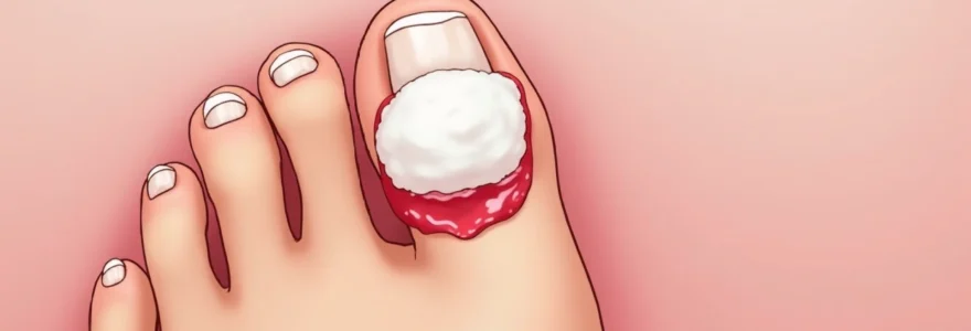
Immediate first aid protocol for lifted toenail management
Prompt first aid intervention following nail injury or recognition of onycholysis significantly influences healing outcomes and complication rates. The primary objectives include bleeding control, infection prevention, pain management, and stabilisation of any remaining attached nail structures. Initial assessment should determine the extent of injury, presence of foreign debris, and signs of underlying tissue damage requiring professional medical attention.
Emergency care begins with thorough hand hygiene and preparation of a clean working environment. Visual inspection helps identify the severity of detachment, presence of active bleeding, and any obvious contamination. Immediate pain relief can be achieved through elevation of the affected foot and application of ice wrapped in a clean cloth for 10-15 minute intervals.
Sterile saline irrigation technique using 0.9% sodium chloride
Sterile saline irrigation represents the gold standard for initial wound cleansing in nail injuries, providing effective debris removal without tissue damage. The isotonic nature of 0.9% sodium chloride solution ensures compatibility with human tissues while maintaining adequate cleansing properties. Irrigation should be performed using gentle pressure to avoid forcing debris deeper into wound spaces or causing additional tissue trauma.
The technique involves directing saline flow from proximal to distal areas, allowing gravity to assist in debris removal. Multiple irrigation cycles may be necessary for heavily contaminated wounds, with each cycle using fresh sterile solution. Avoid using hydrogen peroxide or alcohol-based solutions directly on exposed nail bed tissues, as these can cause additional cellular damage and impair healing processes.
Haemostasis control with direct pressure and elevation methods
Bleeding control in nail injuries typically responds well to direct pressure application using clean, absorbent materials. Gauze pads or clean cloth provide optimal pressure distribution while absorbing blood and wound exudate. Pressure should be applied steadily for 10-15 minutes without frequent checking, as this disrupts clot formation and prolongs bleeding times.
Elevation of the affected extremity above heart level enhances venous return and reduces arterial pressure to the injury site. This positioning proves particularly effective when combined with direct pressure application. Persistent bleeding beyond 20 minutes of appropriate pressure and elevation indicates potential arterial involvement requiring immediate medical evaluation.
Antiseptic application: Povidone-Iodine vs chlorhexidine solutions
Antiseptic selection for nail bed injuries requires balancing antimicrobial efficacy with tissue compatibility and healing promotion. Povidone-iodine provides broad-spectrum antimicrobial activity against bacteria, fungi, and viruses, with minimal tissue toxicity when used in appropriate concentrations. The 10% solution should be diluted to 1% for wound application to prevent tissue irritation while maintaining antimicrobial effectiveness.
Chlorhexidine gluconate offers superior residual antimicrobial activity compared to povidone-iodine, with studies demonstrating continued effectiveness for up to 6 hours post-application. However, chlorhexidine may cause tissue staining and has limited antifungal activity compared to iodine-based preparations. Patient allergies to either agent necessitate alternative antiseptic choices such as dilute sodium hypochlorite solutions.
Non-adherent dressing selection: jelonet and mepitel comparisons
Non-adherent dressing selection proves crucial for nail bed injuries, as standard gauze materials can adhere to wound surfaces and cause re-traumatisation during dressing changes. Jelonet paraffin gauze provides a cost-effective option with good non-adherent properties and moisture retention capabilities. The paraffin impregnation prevents tissue adherence while allowing wound exudate passage through the mesh structure.
Mepitel silicone dressings offer superior non-adherent properties with gentle removal characteristics that minimise patient discomfort and tissue trauma. The silicone coating maintains its non-adherent properties even in the presence of wound exudate, making it ideal for highly exudative nail bed injuries. Transparent variants allow visual wound monitoring without dressing removal, reducing infection risks and patient discomfort.
Professional medical interventions and surgical options
Professional medical intervention becomes necessary when conservative management proves insufficient or when complications develop during the healing process. Podiatrists and dermatologists possess specialised expertise in nail disorders, offering advanced diagnostic capabilities and treatment modalities not available through general medical practitioners. The decision for professional intervention depends on factors including infection presence, extent of nail involvement, underlying medical conditions, and patient comfort with self-management.
Comprehensive professional assessment includes detailed medical history, physical examination of the affected nail and surrounding tissues, and potentially diagnostic testing to identify underlying causes. Dermoscopy may reveal subtle changes in nail bed vasculature and structure not visible through standard examination. Microbiological sampling helps identify specific pathogens when infection is suspected, guiding targeted antimicrobial therapy.
Nail bed debridement using curettes and iris scissors
Nail bed debridement removes devitalised tissue, debris, and infected material that impedes healing and promotes bacterial proliferation. The procedure requires local anaesthesia to ensure patient comfort and enable thorough cleansing of affected areas. Curettes provide controlled tissue removal with minimal damage to healthy nail bed structures, while iris scissors allow precise excision of loose nail plate fragments.
The debridement process begins with careful identification of viable versus non-viable tissues, with healthy nail bed appearing pink and well-vascularised. Gentle curettage removes hyperkeratotic debris and inflammatory exudate while preserving essential nail bed architecture. Complete haemostasis following debridement prevents haematoma formation and optimises healing conditions.
Partial matricectomy for chronic ingrown complications
Partial matricectomy addresses chronic nail problems involving the nail matrix, particularly when onycholysis combines with ingrown nail complications. This surgical technique involves targeted destruction of specific nail matrix areas to prevent problematic nail regrowth while preserving normal nail production in unaffected regions. The procedure requires precise anatomical knowledge and specialised surgical skills to achieve optimal outcomes.
Patient selection for partial matricectomy considers factors including previous conservative treatment failures, recurrent infections, and patient lifestyle requirements. Post-operative care protocols include regular dressing changes, antibiotic prophylaxis in selected cases, and monitoring for complications such as infection or excessive scar tissue formation.
Phenol chemical cauterisation technique for permanent results
Phenol chemical cauterisation provides permanent destruction of targeted nail matrix areas through controlled chemical burns. The 89% phenol solution creates precise tissue destruction when applied with appropriate technique and timing. This method offers advantages including reduced bleeding, decreased post-operative pain, and reliable permanent results when performed correctly.
The procedure requires meticulous preparation including thorough digit cleansing, tourniquet application for haemostasis, and protection of surrounding tissues from inadvertent phenol contact. Neutralisation with alcohol following appropriate exposure time prevents excessive tissue destruction and optimises healing outcomes. Success rates exceed 95% when proper technique and patient selection criteria are followed.
Laser ablation treatment with CO2 and diode technologies
Laser ablation represents the latest advancement in nail matrix destruction, offering precise tissue targeting with minimal collateral damage. CO2 lasers provide excellent tissue vaporisation capabilities with good haemostatic properties, while diode lasers offer deeper tissue penetration and improved post-operative comfort. Both technologies require specialised training and equipment but deliver superior cosmetic outcomes compared to traditional methods.
The laser procedure allows real-time visualisation of tissue destruction, enabling precise control over treatment extent and depth. Reduced post-operative morbidity compared to chemical or surgical techniques makes laser ablation particularly attractive for patients requiring rapid return to normal activities. However, higher equipment costs and specialised training requirements limit widespread availability.
Advanced wound care management and healing optimisation
Advanced wound care protocols for onycholysis focus on creating optimal healing environments while addressing specific challenges associated with nail bed injuries. The unique anatomy and physiology of nail structures require specialised approaches that differ significantly from standard wound management principles. Nail beds possess limited blood supply and depend heavily on diffusion for nutrient delivery, making them particularly susceptible to healing complications when standard protocols are applied inappropriately.
Modern wound care emphasises moisture balance maintenance, bacterial load management, and protection from mechanical trauma during the healing process. Moist wound healing principles apply to nail bed injuries, with studies demonstrating faster epithelialisation and improved cosmetic outcomes when appropriate moisture levels are maintained. However, excessive moisture can promote bacterial overgrowth and maceration, requiring careful balance between hydration and infection prevention.
Advanced dressing technologies incorporating antimicrobial agents, growth factors, and bioactive compounds offer significant advantages over traditional gauze-based approaches in nail bed wound management.
Negative pressure wound therapy has shown promising results in complex nail bed injuries, particularly when combined with traditional onycholysis. The controlled suction removes excess exudate while promoting granulation tissue formation and reducing bacterial contamination. This approach proves particularly valuable in diabetic patients or those with compromised healing capabilities due to vascular insufficiency or immunosuppression.
Biofilm disruption represents a critical component of advanced wound care in onycholysis cases, as bacterial communities within biofilms demonstrate increased resistance to standard antimicrobial treatments. Ultrasonic debridement and specialised antimicrobial dressings help disrupt established biofilms while preventing reformation during the healing process. Regular assessment and modification of treatment protocols based on wound response ensures optimal outcomes and prevents chronic wound development.
Preventing recurrent onycholysis through biomechanical correction
Biomechanical factors play a crucial role in onycholysis development and recurrence, with foot structure abnormalities and gait patterns contributing significantly to repetitive nail trauma. Comprehensive biomechanical assessment identifies specific risk factors including toe deformities, pressure distribution abnormalities, and dynamic foot function issues that predispose individuals to recurrent nail problems. Addressing these underlying mechanical factors proves essential for long-term prevention of onycholysis recurrence.
Orthotic therapy provides effective biomechanical correction through redistribution of plantar pressures and improvement of foot alignment during weight-bearing activities. Custom-fabricated orthotics offer superior outcomes compared to over-the-counter alternatives, with specific modifications targeting individual patient needs and foot structure variations. Pressure relief over affected toe areas prevents repetitive trauma while accommodating any residual nail deformities resulting from previous onycholysis episodes.
Proper footwear selection and modification represent fundamental components of onycholysis prevention, with studies indicating that inappropriate shoe fit contributes to over 60% of recurrent cases.
Gait analysis reveals dynamic factors contributing to excessive toe pressures during walking and running activities. High-speed video analysis and pressure mapping systems identify specific phases of gait cycle where peak pressures occur over vulnerable nail areas. Gait retraining programmes help patients modify harmful movement patterns while maintaining functional mobility and athletic performance when applicable.
Activity modification recommendations balance prevention requirements with patient lifestyle goals and occupational demands. Athletes may require sport-specific interventions including protective padding, modified training schedules, and alternative footwear options during high-risk activities. Occupational considerations include workplace safety requirements, prolonged standing demands, and exposure to chemical or moisture hazards that may compromise nail health.
Long-term nail regrowth monitoring and complications management
Long-term monitoring of nail regrowth following onycholysis requires systematic assessment protocols that track healing progress while identifying potential complications early in their development. The nail regrowth process spans 12-18 months for toenails, during which multiple factors can influence final outcomes including infection development, mechanical trauma, and underlying systemic conditions. Regular monitoring appointments allow for timely intervention when complications arise and optimisation
of healing parameters through evidence-based interventions.Initial regrowth typically becomes visible 4-6 weeks following complete nail loss, appearing as a thin, translucent structure emerging from the proximal nail fold. This early growth phase requires protection from mechanical trauma and maintenance of optimal moisture conditions to prevent structural abnormalities. Growth rate variations depend on patient age, nutritional status, circulation quality, and presence of underlying medical conditions affecting cellular metabolism.Monthly photographic documentation provides objective assessment of regrowth progress and enables early detection of abnormal growth patterns. Digital imaging with standardised lighting and positioning allows accurate measurement of growth rates and identification of surface irregularities or colour changes that may indicate developing complications. Comparison with baseline images helps track improvement trends and guides treatment modifications when progress deviates from expected patterns.The quality of nail regrowth depends significantly on preservation of nail matrix integrity during the acute injury phase. Damage to matrix cells results in permanent structural abnormalities including ridging, thinning, or irregular surface texture that persists throughout the nail’s lifetime. Matrix scarring from severe infections or inappropriate treatment techniques commonly produces dystrophic nail growth requiring ongoing management and patient education about long-term implications.Complications during regrowth include recurrent infection development, ingrown nail formation, and adherence problems between new nail plate and underlying nail bed. Secondary bacterial infections frequently occur when protective barriers fail or when patients resume normal activities too early in the healing process. Early recognition of infection signs including increased pain, erythema, purulent discharge, or foul odour enables prompt antibiotic intervention before systemic spread occurs.
Regular monitoring appointments scheduled at 4-6 week intervals during the first six months of regrowth allow healthcare providers to identify complications early and adjust treatment protocols accordingly.
Ingrown nail development represents a common complication following onycholysis resolution, particularly when original injury involved lateral nail fold trauma or when biomechanical factors remain uncorrected. The combination of altered nail growth patterns and residual inflammation creates conditions favouring lateral nail penetration into surrounding soft tissues. Prophylactic lateral nail fold protection using specialised padding or splinting techniques prevents ingrown complications while allowing normal growth progression.Adherence problems between regenerating nail plate and nail bed occur most frequently in cases where extensive nail bed damage occurred during the initial injury or treatment phase. Poor adherence results in recurrent lifting episodes and creates ongoing risks for infection development and chronic discomfort. These cases may require specialised interventions including nail bed grafting procedures or permanent partial nail removal to achieve acceptable functional outcomes.Patient education regarding realistic expectations for regrowth outcomes proves essential for psychological adjustment and treatment compliance. Complete restoration of pre-injury nail appearance occurs in only 60-70% of cases, with remaining patients experiencing some degree of permanent cosmetic or functional impairment. Counselling support helps patients adapt to altered nail appearance and develop coping strategies for ongoing management requirements.Long-term follow-up protocols extend beyond complete nail regrowth to monitor for late complications including recurrent onycholysis, chronic pain syndromes, or development of secondary skin conditions around affected digits. Annual assessment appointments allow early intervention for emerging problems while providing ongoing education about prevention strategies and self-monitoring techniques. The comprehensive approach to onycholysis management encompasses immediate treatment, healing optimisation, and long-term prevention strategies that address both local nail pathology and underlying contributing factors.