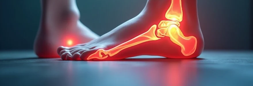
Experiencing persistent pain three months following cheilectomy surgery raises legitimate concerns for patients who expected to be well on their road to recovery by this stage. While some degree of discomfort during the healing process is anticipated, understanding what constitutes normal versus problematic pain patterns becomes crucial for proper treatment outcomes. The complexity of foot surgery recovery, particularly involving the metatarsophalangeal joint, means that healing timelines can vary significantly between individuals.
Cheilectomy, designed to address hallux rigidus by removing bone spurs and damaged tissue from the big toe joint, typically follows a predictable recovery pattern. However, various factors including surgical technique, individual healing capacity, and post-operative care adherence can influence pain levels and duration. Distinguishing between expected healing discomfort and pathological pain requiring medical attention becomes essential for optimal patient outcomes.
Understanding normal cheilectomy recovery timeline and pain patterns
The recovery process following cheilectomy surgery follows distinct phases, each characterised by specific pain patterns and healing milestones. During the initial post-operative period, patients typically experience significant pain that gradually diminishes over the first several weeks. However, the three-month mark represents a critical assessment point where pain levels should have substantially decreased from their immediate post-surgical intensity.
Expected pain trajectory during first three months Post-Surgery
Most patients can expect pain levels to follow a predictable downward trajectory following cheilectomy. The first 48-72 hours typically involve the most intense discomfort, requiring prescription pain medication for adequate management. By the two-week mark, when sutures are removed, pain should have reduced to manageable levels allowing for gradual return to weight-bearing activities. The six-week period usually marks significant improvement, with many patients reporting substantial pain relief.
At three months post-surgery, residual pain should be minimal and primarily related to increased activity levels or weather changes. Persistent moderate to severe pain at this stage warrants careful evaluation to determine underlying causes. Normal healing pain at three months typically presents as mild aching or stiffness rather than sharp, constant, or debilitating discomfort.
Distinguishing between inflammatory pain and mechanical discomfort
Understanding the difference between inflammatory and mechanical pain sources helps patients assess whether their three-month post-operative discomfort falls within normal parameters. Inflammatory pain often presents with accompanying swelling, warmth, and redness around the surgical site. This type of pain typically responds well to anti-inflammatory medications and rest, gradually improving with time.
Mechanical discomfort, conversely, relates to joint movement and weight-bearing activities. Patients may experience increased pain with prolonged standing or walking, which improves with rest. This mechanical pain should progressively decrease as the joint adapts to its new biomechanics following bone spur removal. Persistent mechanical pain beyond three months may indicate incomplete bone healing or joint stiffness requiring intervention.
Bone remodelling process and associated sensations
The bone remodelling process continues well beyond the initial healing period, potentially causing intermittent discomfort up to six months post-surgery. During cheilectomy, bone surfaces are modified through precise removal of osteophytes and joint debris. The remaining bone tissue undergoes remodelling to adapt to altered mechanical stresses and improve joint function.
Patients may experience occasional aching or throbbing sensations as new bone formation occurs and existing bone structure adapts. These sensations often intensify with increased activity levels or changes in barometric pressure. The remodelling process typically stabilises by six months , with most patients experiencing significant improvement in comfort and function by this timeframe.
Soft tissue healing phases following metatarsophalangeal joint surgery
Soft tissue healing around the metatarsophalangeal joint involves multiple structures including skin, subcutaneous tissue, joint capsule, and surrounding tendons. The initial inflammatory phase lasts approximately 2-3 days, followed by the proliferative phase extending through several weeks. The maturation phase, during which tissue strength improves and scar formation stabilises, continues for several months.
At three months post-cheilectomy, soft tissue healing should be largely complete, though final maturation may continue. Patients might experience occasional tightness or pulling sensations as scar tissue continues to remodel. Persistent swelling or soft tissue tenderness beyond three months may indicate delayed healing or complications requiring medical evaluation.
Pathological pain indicators three months after cheilectomy
Recognising pathological pain patterns three months following cheilectomy enables early identification and management of complications that could compromise surgical outcomes. While some degree of discomfort may persist during normal healing, certain pain characteristics suggest underlying problems requiring immediate medical attention. Understanding these warning signs empowers patients to seek appropriate care when recovery deviates from expected patterns.
Complex regional pain syndrome development following foot surgery
Complex Regional Pain Syndrome (CRPS) represents a serious complication that can develop following any foot surgery, including cheilectomy. This condition typically manifests within the first few months after surgery and presents with disproportionate pain compared to the surgical procedure performed. Patients with CRPS often describe burning, crushing, or shooting pain that extends beyond the surgical site.
Additional symptoms accompanying CRPS include skin colour changes, temperature variations, and increased sensitivity to touch or light pressure. The affected foot may appear shiny, swollen, or show abnormal hair and nail growth patterns. Early recognition and treatment of CRPS significantly improves patient outcomes , making prompt medical evaluation essential when these symptoms arise. Treatment typically involves a multidisciplinary approach including pain management specialists, physiotherapy, and sometimes sympathetic nerve blocks.
Infection-related pain patterns and diagnostic markers
While surgical site infections typically manifest within the first few weeks following cheilectomy, delayed infections can occasionally occur up to several months post-operatively. Infection-related pain often presents as constant, throbbing discomfort that progressively worsens rather than improving over time. This pain pattern contrasts sharply with normal healing pain, which generally follows a decreasing trajectory.
Accompanying signs of infection include persistent swelling, warmth, redness around the incision site, and potential drainage or wound breakdown. Patients may also experience systemic symptoms such as fever, chills, or general malaise. Deep infections affecting bone or joint structures require immediate antibiotic treatment and may necessitate surgical intervention to prevent long-term complications. Blood tests showing elevated inflammatory markers support the diagnosis of infection.
Hardware-related complications and screw loosening symptoms
Although traditional cheilectomy typically doesn’t require internal fixation hardware, some surgical variations or concurrent procedures may involve screws or other implants. When hardware is present, loosening or migration can cause persistent pain that often worsens with activity. Patients may describe a grinding sensation or mechanical catching within the joint during movement.
Hardware-related complications can also include irritation of surrounding soft tissues, leading to localised pain and swelling. Imaging studies such as X-rays or CT scans help identify hardware positioning and potential complications. Symptomatic hardware problems may require revision surgery to remove or reposition problematic implants, particularly when conservative management fails to provide adequate relief.
Recurrent hallux rigidus and progressive joint degeneration signs
Unfortunately, some patients may experience recurrence of hallux rigidus symptoms following cheilectomy, particularly if underlying joint disease continues to progress. Recurrent bone spur formation or progressive joint space narrowing can cause renewed pain and stiffness. This pain often resembles the original symptoms that prompted surgery, including difficulty with toe dorsiflexion and pain during push-off phase of walking.
Progressive joint degeneration may indicate that the initial surgical intervention was insufficient for the degree of arthritis present, or that the condition has continued to advance despite treatment. Patients typically notice gradual return of stiffness and pain that initially improves with rest but progressively becomes more constant. Advanced imaging studies can reveal the extent of joint degeneration and help guide future treatment decisions, which may include more extensive surgical interventions such as joint fusion or replacement.
Clinical assessment methods for persistent Post-Cheilectomy pain
Comprehensive clinical assessment of persistent pain three months following cheilectomy requires systematic evaluation combining detailed history taking, physical examination, and appropriate diagnostic imaging. Healthcare providers must differentiate between normal healing variants and pathological processes that may compromise long-term outcomes. The assessment process begins with characterising pain patterns, including onset, duration, intensity, and aggravating or relieving factors.
Physical examination focuses on evaluating wound healing status, joint range of motion, and functional capacity compared to pre-operative baselines. Clinicians assess for signs of infection, including localised warmth, erythema, or drainage, while also examining overall foot biomechanics and gait patterns. Systematic palpation of anatomical structures helps localise pain sources and identify potential complications such as nerve irritation or soft tissue adhesions.
Diagnostic imaging plays a crucial role in identifying structural abnormalities contributing to persistent pain. Standard weight-bearing radiographs reveal bone healing status, joint alignment, and potential hardware complications when applicable. Advanced imaging modalities such as magnetic resonance imaging (MRI) provide detailed soft tissue evaluation, identifying potential sources of pain including joint effusion, soft tissue inflammation, or nerve compression. Bone scintigraphy may occasionally be employed to assess bone metabolism and identify areas of increased activity suggesting ongoing pathological processes.
Laboratory studies complement imaging findings when infection or systemic inflammatory conditions are suspected. Elevated inflammatory markers including C-reactive protein and erythrocyte sedimentation rate may indicate ongoing infectious or inflammatory processes. Complete blood count and differential can reveal signs of systemic infection or other haematological abnormalities that might influence healing. Joint aspiration with synovial fluid analysis becomes necessary when septic arthritis is suspected, providing definitive diagnostic information and guiding antibiotic selection.
Conservative management strategies for extended recovery periods
Conservative management approaches for persistent pain three months post-cheilectomy focus on optimising healing conditions while addressing specific pain mechanisms that may be prolonging recovery. Anti-inflammatory medications remain a cornerstone of treatment, helping reduce residual inflammation that may be contributing to ongoing discomfort. Topical preparations may provide localised relief while minimising systemic side effects, particularly beneficial for patients with contraindications to oral anti-inflammatory drugs.
Physical therapy interventions target joint mobility, soft tissue flexibility, and functional restoration through graduated exercise programmes. Manual therapy techniques including joint mobilisation and soft tissue manipulation help address adhesions and stiffness that commonly develop following foot surgery. Progressive strengthening exercises restore muscle function and improve overall foot biomechanics, reducing compensatory movement patterns that may perpetuate pain. Gait training ensures proper weight distribution and movement patterns during walking activities.
Orthotic interventions provide external support and modify foot biomechanics to reduce stress on healing tissues. Custom or over-the-counter orthotic devices can redistribute pressure away from the surgical site while promoting proper foot alignment during weight-bearing activities. Shoe modifications, including rocker bottom soles or increased toe box height, accommodate post-surgical changes while facilitating normal gait patterns. Proper footwear selection becomes crucial for long-term comfort and function following cheilectomy surgery.
Pain management techniques encompass both pharmacological and non-pharmacological approaches tailored to individual patient needs. Prescription pain medications may be necessary for patients experiencing significant discomfort, though long-term use requires careful monitoring to prevent dependency. Alternative pain management strategies including transcutaneous electrical nerve stimulation (TENS), acupuncture, and mindfulness-based interventions provide additional options for patients seeking non-pharmaceutical approaches. Heat and cold therapy applications can provide symptomatic relief while potentially facilitating the healing process.
Surgical revision considerations and alternative treatment approaches
Surgical revision becomes a consideration when conservative management fails to provide adequate pain relief three months following cheilectomy, particularly when imaging studies reveal specific structural abnormalities amenable to surgical correction. The decision-making process requires careful evaluation of potential benefits versus risks, considering patient factors including overall health status, functional goals, and previous surgical outcomes. Revision procedures carry inherently higher complication rates compared to primary interventions, necessitating thorough patient counselling regarding expected outcomes.
Arthroscopic evaluation and treatment represent minimally invasive options for addressing persistent pain when the underlying cause remains unclear despite comprehensive diagnostic evaluation. Arthroscopic techniques allow direct visualisation of intra-articular structures while enabling therapeutic interventions such as synovectomy, loose body removal, or chondral debridement. This approach provides both diagnostic and therapeutic benefits while minimising additional surgical trauma to already compromised tissues. Recovery from arthroscopic procedures typically involves shorter rehabilitation periods compared to open revision surgery.
More extensive surgical interventions may be necessary when significant joint degeneration or recurrent bone spur formation contributes to persistent symptoms. Joint fusion (arthrodesis) eliminates painful joint motion while providing stability and pain relief for patients with advanced arthritis unresponsive to joint-preserving procedures. Joint replacement technologies continue to evolve , offering motion-preserving alternatives for select patients, though long-term outcomes data remains limited for metatarsophalangeal joint replacements compared to other joint replacement procedures.
Alternative treatment approaches encompass regenerative medicine techniques that may promote healing and reduce pain without requiring additional surgical intervention. Platelet-rich plasma injections utilise concentrated growth factors from the patient’s own blood to potentially accelerate healing and reduce inflammation. Stem cell therapies, while still investigational, show promise for cartilage regeneration and pain reduction in joint disorders. These biological treatments may serve as bridge therapies while patients continue conservative management or as alternatives for patients who are poor surgical candidates due to medical comorbidities.