White patches, spots, or streaks on fingernails and toenails represent one of the most frequently encountered nail abnormalities in clinical practice. These distinctive markings, medically termed leukonychia, can manifest in various forms ranging from tiny punctate spots to extensive bands traversing the entire nail plate. While most instances prove harmless and temporary, understanding the underlying mechanisms and diverse aetiologies remains crucial for both healthcare professionals and individuals experiencing these nail changes.
The prevalence of nail leukonychia varies considerably across different populations, with studies indicating that up to 90% of individuals will experience some form of white nail discolouration during their lifetime. These manifestations can occur at any age, though certain patterns demonstrate distinct demographic preferences. The condition’s impact extends beyond mere cosmetic concerns, as specific forms may signal underlying systemic health conditions requiring immediate medical attention.
Leukonychia: understanding white nail discolouration pathophysiology
Leukonychia encompasses a spectrum of nail plate abnormalities characterised by partial or complete loss of normal nail transparency. The pathophysiological mechanisms underlying white nail discolouration involve complex interactions between keratinocyte disruption, altered nail matrix function, and various external or internal factors affecting nail plate formation. Understanding these fundamental processes provides essential insight into both the development and clinical significance of different leukonychia presentations.
The nail plate consists predominantly of keratin fibres arranged in distinct layers, with normal nail transparency dependent upon proper keratinocyte alignment and hydration levels. When this organised structure becomes disrupted, light scattering increases significantly, resulting in the characteristic opaque white appearance. This disruption can occur at multiple levels within the nail formation process, from initial matrix dysfunction to post-formation trauma or chemical exposure.
Classification systems for leukonychia consider both anatomical distribution patterns and underlying pathophysiological mechanisms. True leukonychia originates within the nail matrix itself, whilst apparent leukonychia results from changes in the underlying nail bed or surrounding tissues. This distinction proves clinically significant , as true leukonychia typically grows out with normal nail growth, whereas apparent forms may persist or fluctuate based on the underlying condition’s progression.
Punctate leukonychia: keratinocyte disruption mechanisms
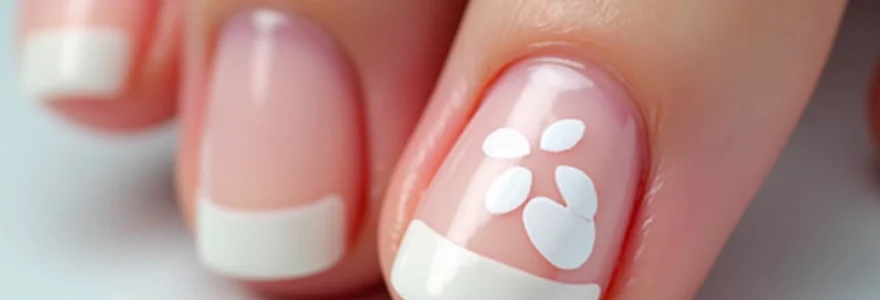
Punctate leukonychia represents the most common form of white nail discolouration, characterised by small, discrete white spots scattered across the nail plate surface. These lesions typically measure 1-3 millimetres in diameter and result from localised keratinocyte disruption within specific areas of the nail matrix. The precise mechanisms involve temporary cessation of normal keratin synthesis, leading to air pocket formation within the affected nail plate regions.
Recent research has demonstrated that punctate lesions develop following minor trauma to the proximal nail fold or matrix area, often occurring days or weeks before visible manifestation. The delayed appearance reflects the time required for affected nail plate areas to migrate distally as normal nail growth progresses. This temporal relationship frequently complicates accurate cause identification, as patients may not recall the initiating traumatic event.
Striate leukonychia: linear white band formation
Striate or transverse leukonychia manifests as horizontal white lines extending across the nail plate width, parallel to the lunula. These distinctive bands, sometimes termed Mees’ lines, typically indicate more significant or prolonged matrix disruption compared to punctate lesions. The linear pattern suggests that the causative factor affected the entire nail matrix simultaneously, resulting in uniform abnormality across the nail plate width.
The formation of striate leukonychia often correlates with systemic illnesses, metabolic disturbances, or toxic exposures that temporarily impair nail matrix function. The band width directly relates to the duration of matrix dysfunction , with broader lines indicating more prolonged disruption periods. Multiple parallel bands may develop when repeated episodes of matrix dysfunction occur during the nail growth cycle.
Total leukonychia: complete nail plate opacity
Total leukonychia presents as complete nail plate whitening, affecting the entire visible nail surface from lunula to free edge. This dramatic presentation typically indicates severe or chronic matrix dysfunction, often associated with congenital conditions or significant systemic diseases. The complete opacity results from extensive keratinocyte disruption throughout the nail formation process, preventing normal nail plate transparency.
Acquired total leukonychia may develop gradually over months or years, particularly in association with chronic kidney disease, liver dysfunction, or severe protein malnutrition. The condition often proves irreversible once established, requiring ongoing management of underlying systemic conditions to prevent progression to additional nails.
Partial leukonychia: localised white patch development
Partial leukonychia encompasses various patterns of incomplete nail plate whitening, ranging from small localised patches to extensive areas affecting significant nail plate portions. These manifestations can occur anywhere on the nail surface and may exhibit regular or irregular borders depending on the underlying cause. The heterogeneous presentation reflects the diverse aetiologies capable of producing partial nail plate opacity.
Longitudinal leukonychia represents a specific subset characterised by white bands extending parallel to nail growth direction. This pattern typically indicates localised matrix dysfunction affecting specific areas over extended periods.
The longitudinal pattern suggests ongoing or recurrent matrix disruption in discrete anatomical locations, often associated with chronic trauma or localised inflammatory conditions.
Traumatic nail matrix injury and white spot formation
Trauma represents the predominant cause of nail leukonychia, accounting for approximately 70-80% of cases presenting to dermatology clinics. The nail matrix demonstrates exquisite sensitivity to mechanical disruption, with even minor trauma capable of producing visible white spot formation. Understanding the various traumatic mechanisms and their characteristic presentations enables accurate diagnosis and appropriate patient counselling regarding prognosis and prevention strategies.
The temporal relationship between trauma and visible leukonychia manifestation varies considerably based on nail growth rates and the anatomical location of matrix injury. Fingernails typically require 4-6 months for complete regeneration, whilst toenails may take 12-18 months. Consequently, white spots appearing on distal nail areas may represent trauma occurring many months previously , complicating accurate cause identification in clinical practice.
Matrix sensitivity varies significantly between individuals and may be influenced by factors such as age, nutritional status, and concurrent medical conditions. Children often demonstrate increased susceptibility to traumatic leukonychia due to thinner nail plates and higher likelihood of engaging in activities resulting in nail trauma. Conversely, elderly individuals may experience reduced matrix sensitivity, potentially masking mild traumatic episodes.
Mechanical trauma from manicure tools and cuticle manipulation
Professional and home manicure procedures represent significant sources of nail matrix trauma, particularly when aggressive cuticle manipulation or excessive filing occurs. Modern electric nail files and cuticle nippers can generate substantial mechanical forces, easily exceeding the matrix’s tolerance for external pressure. The proximal nail fold area demonstrates particular vulnerability, as aggressive pushing or cutting can directly damage underlying matrix structures.
Gel and acrylic nail application procedures frequently involve extensive nail surface preparation, including aggressive buffing and chemical treatments that may compromise nail plate integrity. The removal process presents additional trauma risks, particularly when improper techniques involving forceful scraping or excessive chemical exposure are employed. Regular professional manicures using proper techniques significantly reduce trauma-associated leukonychia risks whilst maintaining optimal nail health.
Repetitive stress injuries from nail biting and picking
Nail biting and picking behaviours, collectively termed onychophagia, represent common sources of chronic nail matrix trauma. These repetitive behaviours often target the proximal nail fold and surrounding tissues, leading to persistent low-grade inflammation and matrix disruption. The chronic nature of these activities frequently results in multiple white spots appearing sequentially as damaged nail areas migrate distally with normal growth.
The psychological aspects of nail biting and picking often complicate treatment approaches, as addressing the underlying behavioural patterns proves essential for preventing recurrent leukonychia. Stress, anxiety, and obsessive-compulsive tendencies frequently contribute to these behaviours, necessitating comprehensive management strategies addressing both the nail pathology and underlying psychological factors.
Onychotillomania: compulsive nail manipulation effects
Onychotillomania represents a severe form of compulsive nail manipulation characterised by repetitive picking, biting, or scratching behaviours targeting the nail apparatus. This condition often results in extensive nail damage, including multiple forms of leukonychia, nail plate dystrophy, and secondary bacterial infections. The compulsive nature of these behaviours frequently leads to significant functional impairment and social embarrassment.
Treatment approaches for onychotillomania-associated leukonychia must address both the underlying psychiatric condition and resultant nail pathology.
Successful management typically requires multidisciplinary collaboration between dermatologists, psychiatrists, and behavioural therapists to achieve optimal outcomes and prevent recurrence.
Pharmacological interventions may include selective serotonin reuptake inhibitors or other psychiatric medications alongside topical nail treatments.
Sports-related nail bed trauma and recovery patterns
Athletic activities present unique risks for nail trauma, particularly in sports involving repetitive impact or pressure on the digits. Running sports frequently cause toenail trauma from repetitive shoe impact, whilst racquet sports may produce fingernail injuries from equipment vibration or ball contact. The specific patterns of sports-related leukonychia often reflect the characteristic movement patterns and equipment use associated with particular athletic activities.
Recovery patterns following sports-related nail trauma typically demonstrate good prognosis when appropriate protective measures are implemented. Proper footwear fitting, protective gloves or tape application, and technique modification can significantly reduce trauma recurrence risks. Athletes experiencing recurrent nail leukonychia should undergo comprehensive biomechanical assessment to identify and address contributing factors.
Systemic medical conditions manifesting as nail leukonychia
Systemic diseases affecting multiple organ systems frequently manifest with characteristic nail changes, including various forms of leukonychia. These presentations often provide valuable diagnostic clues, particularly when other clinical signs remain subtle or absent. Recognition of nail manifestations associated with systemic conditions enables earlier diagnosis and treatment initiation, potentially improving overall patient outcomes significantly.
The mechanisms underlying systemic disease-associated leukonychia involve complex interactions between altered metabolism, protein synthesis disruption, and compromised tissue perfusion. Chronic kidney disease, liver dysfunction, and severe malnutrition represent the most common systemic causes, though numerous other conditions may produce similar nail manifestations. The specific leukonychia patterns often correlate with disease severity and duration, providing prognostic information regarding underlying condition progression.
Apparent leukonychia associated with systemic diseases typically demonstrates characteristic features distinguishing it from traumatic causes. These include bilateral symmetrical involvement, concurrent nail abnormalities such as clubbing or koilonychia, and associated systemic symptoms. The white discolouration often fluctuates with disease activity , becoming more prominent during exacerbations and improving with effective treatment of the underlying condition.
Hypoalbuminaemia and protein deficiency indicators
Severe hypoalbuminaemia, typically defined as serum albumin levels below 2.5 g/dL, frequently produces characteristic nail changes including transverse white bands and apparent leukonychia. The pathophysiological mechanism involves altered nail bed perfusion and reduced protein availability for normal keratin synthesis. These changes often appear when albumin levels remain depressed for extended periods, typically several weeks to months.
Muehrcke’s lines represent a specific form of apparent leukonychia characterised by paired transverse white bands separated by normal nail colour. These distinctive markings occur exclusively in association with severe hypoalbuminaemia and typically disappear when albumin levels normalise. The bands do not move with nail growth, distinguishing them from true leukonychia associated with matrix dysfunction.
Chronic kidney disease and uraemic nail changes
Advanced chronic kidney disease frequently produces characteristic nail abnormalities, including apparent leukonychia, nail plate thinning, and altered growth patterns. The pathophysiological mechanisms involve uraemic toxin accumulation, altered mineral metabolism, and chronic anaemia affecting nail bed vasculature. These changes often correlate with disease severity and may improve following successful renal replacement therapy or transplantation.
Half-and-half nails, also termed Lindsay’s nails, represent a specific pattern associated with chronic kidney disease characterised by white proximal nail areas and reddish-brown distal bands. This distinctive presentation results from altered nail bed vasculature and chronic uraemic effects on nail matrix function. The appearance often correlates with estimated glomerular filtration rate , providing a visual indicator of renal function deterioration or improvement.
Liver cirrhosis and terry’s nails presentation
Advanced liver cirrhosis frequently manifests with Terry’s nails, characterised by white nail plates with narrow pink bands at the distal nail edge. This distinctive presentation results from altered albumin synthesis, portal hypertension effects on nail bed circulation, and chronic liver dysfunction impacts on overall nail metabolism. The condition typically affects multiple nails simultaneously and often proves irreversible despite liver function improvement.
The pathophysiology underlying Terry’s nails involves complex interactions between reduced albumin synthesis, altered vascular dynamics, and chronic inflammatory processes associated with liver cirrhosis.
The severity of nail changes often correlates with Child-Pugh classification scores and overall prognosis, making nail examination a valuable component of liver disease assessment.
Additional liver-associated nail changes may include clubbing, koilonychia, and increased nail fragility.
Zinc deficiency and acrodermatitis enteropathica
Severe zinc deficiency, whether acquired or congenital, frequently produces characteristic nail abnormalities including leukonychia, nail plate thinning, and delayed growth. Acrodermatitis enteropathica, a rare autosomal recessive disorder affecting zinc absorption, typically presents with distinctive nail changes alongside characteristic skin lesions and gastrointestinal symptoms. The nail manifestations often appear early in the disease course and may provide initial diagnostic clues.
Acquired zinc deficiency may result from malabsorption syndromes, chronic diarrhoea, or inadequate dietary intake, particularly in elderly or hospitalised patients. The nail changes typically respond well to zinc supplementation, with improvement visible within weeks of treatment initiation. Regular monitoring of zinc levels proves essential in high-risk populations to prevent deficiency-related nail complications.
Fungal and bacterial nail infections causing white patches
Infectious causes of nail leukonychia encompass various fungal and bacterial pathogens capable of producing characteristic white discolouration patterns. White superficial onychomycosis represents the most common infectious cause, typically affecting toenails and presenting as chalky white patches on the nail surface. The condition results from specific dermatophyte species, most commonly Trichophyton mentagrophytes , directly invading the superficial nail plate layers.
The pathophysiology of infectious leukonychia involves direct pathogen invasion of nail keratin structures, resulting in altered light transmission properties and characteristic white opacity. Unlike traumatic causes, infectious leukonychia typically demonstrates progressive expansion and may be associated with nail plate thickening, surface irregularities, and secondary bacterial superinfection. Early diagnosis and appropriate antifungal treatment prove essential for preventing extensive nail destruction and transmission to other nails.
Bacterial nail infections, though less common than fungal causes, may produce similar white discolouration patterns, particularly when associated with Pseudomonas aeruginosa or Staphylococcus aureus . These infections often occur secondary to trauma or chronic moisture exposure and may require systemic antibiotic therapy for successful resolution. The presence of associated inflammation, purulent discharge, or nail fold swelling should raise suspicion for bacterial aetiology.
Diagnosis of infectious leukonychia requires appropriate laboratory testing, including potassium hydroxide preparation, fungal culture, and potentially nail biopsy for definitive pathogen identification. Treatment duration for fungal nail infections typically ranges from 3-6 months , with success rates varying based on pathogen type, infection extent, and patient compliance. Prevention strategies focus on maintaining proper nail hygiene, avoiding prolonged moisture exposure, and using appropriate footwear in high-risk environments.
Congenital nail dystrophies and hereditary leukonychia
Hereditary forms of leukonychia represent rare but clinically significant conditions characterised by genetic mutations affecting nail formation processes. These conditions typically manifest during infancy or early childhood and often demonstrate autosomal dominant inheritance patterns. The molecular mechanisms involve mutations in genes encoding structural proteins essential for normal nail keratin formation, resulting in characteristic white nail appearances that persist throughout life.
Autosomal dominant leukonychia totalis syndrome
Autosomal dominant leukonychia totalis represents an extremely rare genetic condition characterised by complete nail whitening affecting all twenty digits from birth or early infancy. The condition results from mutations in genes encoding keratin proteins, particularly KRT85 and KRT86, which are essential for normal nail plate formation. These genetic alterations disrupt the organised keratin structure within the nail matrix, leading to altered light transmission properties and the characteristic opaque white appearance.
The inheritance pattern follows classic Mendelian genetics, with affected individuals having a 50% probability of transmitting the condition to their offspring. Clinical presentation typically includes thickened, completely white nail plates that grow at normal rates but never develop normal transparency. The condition remains stable throughout life, with no reported cases of spontaneous improvement or progression to nail destruction. Molecular genetic testing can confirm the diagnosis and provide accurate genetic counselling for affected families.
Associated features may include mild nail plate thickening and increased brittleness, though nail function typically remains preserved. The cosmetic impact often proves significant, particularly during adolescence and young adulthood, necessitating appropriate psychological support and counselling. Current treatment options remain limited to cosmetic camouflage techniques, as the underlying genetic defect cannot be corrected with available therapeutic interventions.
Darier disease and associated nail abnormalities
Darier disease, also known as keratosis follicularis, represents an autosomal dominant genodermatosis caused by mutations in the ATP2A2 gene encoding the sarcoplasmic reticulum calcium pump SERCA2. The condition frequently manifests with characteristic nail abnormalities, including longitudinal white and pink bands, nail plate thinning, and distinctive V-shaped nicking at the free edge. These nail changes often precede the development of characteristic skin lesions and may provide early diagnostic clues.
The pathophysiological mechanism underlying Darier disease nail changes involves disrupted calcium homeostasis within keratinocytes, leading to altered cell adhesion and abnormal keratinisation processes. The longitudinal white bands result from intermittent matrix dysfunction, whilst the characteristic nail nicking reflects structural weakness at specific anatomical locations. Nail changes typically affect multiple digits symmetrically and may worsen during disease exacerbations triggered by heat, friction, or stress.
The distinctive nail abnormalities in Darier disease often provide the first clinical clue to diagnosis, particularly when skin manifestations remain subtle or localised to easily overlooked areas.
Management of Darier disease nail changes focuses on preventing secondary complications and maintaining nail hygiene. Topical retinoids may provide modest improvement in nail appearance, whilst systemic retinoids reserved for severe cases can produce more significant but temporary benefits. Patient education regarding trigger avoidance and proper nail care proves essential for preventing complications and optimising quality of life.
Pachyonychia congenita and nail plate thickening
Pachyonychia congenita encompasses a group of rare autosomal dominant disorders characterised by severe nail dystrophy and associated ectodermal abnormalities. The condition results from mutations in various keratin genes, including KRT6A, KRT6B, KRT6C, KRT16, and KRT17, which are essential for normal nail and skin keratinocyte function. Nail manifestations typically appear during infancy and progress to severe thickening, opacity, and functional impairment throughout childhood and adolescence.
The characteristic nail changes include progressive thickening of all nail plates, often accompanied by white discolouration, increased curvature, and painful ingrowth. The pathophysiology involves disrupted keratin filament organisation within nail matrix cells, leading to abnormal keratinocyte proliferation and differentiation. This results in the production of structurally abnormal nail plates that lack normal flexibility and transparency.
Clinical management of pachyonychia congenita nail changes requires multidisciplinary approach involving dermatologists, podiatrists, and genetic counsellors. Regular nail trimming and filing prove essential for preventing painful impaction and secondary infections. Surgical nail avulsion may be necessary in severe cases, though recurrence with similar abnormalities typically occurs. Gene therapy approaches currently under investigation may offer future therapeutic options for this challenging condition.
Associated features frequently include palmoplantar keratoderma, oral cavity lesions, and various other ectodermal abnormalities that can significantly impact quality of life. Early genetic diagnosis enables appropriate counselling and management planning, whilst patient registries facilitate research into novel therapeutic approaches. The International Pachyonychia Congenita Research Registry continues to collect valuable data regarding natural history and treatment responses in affected individuals worldwide.