Ventral thecal sac effacement represents one of the most significant diagnostic findings encountered in spinal imaging, yet many patients struggle to comprehend its implications when they receive their MRI reports. This condition occurs when structures within the spinal canal compress the protective membrane surrounding the spinal cord from the front, causing it to flatten or indent. The term “effacement” literally means “to wipe out” or “obliterate,” indicating that normal anatomical space is being compromised. Understanding this condition becomes crucial for anyone experiencing symptoms of spinal stenosis, disc herniation, or other degenerative spinal conditions that can lead to neural compression and potentially serious neurological consequences.
Anatomical structure and location of the ventral thecal sac within the spinal column
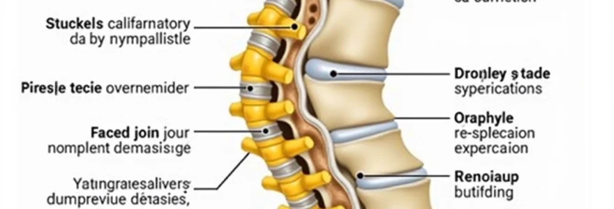
The thecal sac serves as nature’s protective housing for the most critical components of the central nervous system within the spine. This dural membrane structure extends from the foramen magnum at the skull base down to approximately the second sacral vertebra, encasing both the spinal cord and the collection of nerve roots known as the cauda equina. The ventral aspect refers specifically to the front portion of this sac, which faces toward the vertebral bodies and intervertebral discs.
Cerebrospinal fluid circulation patterns around the thecal sac
Within the thecal sac, cerebrospinal fluid continuously circulates, providing essential nutrients to neural tissues while removing metabolic waste products. This fluid circulation follows specific patterns that can become disrupted when ventral effacement occurs. The CSF normally flows freely around the spinal cord, maintaining optimal pressure and creating a cushioning effect that protects delicate neural structures from mechanical stress during daily movements.
Dural membrane composition and protective functions
The dural membrane itself consists of dense fibrous connective tissue that provides robust protection against external forces. This tough outer covering maintains the integrity of the subarachnoid space where cerebrospinal fluid resides. When functioning normally, the dura mater exhibits sufficient flexibility to accommodate normal spinal movements while maintaining its protective barrier function against potentially harmful mechanical compression.
Relationship between thecal sac and adjacent vertebral bodies
The spatial relationship between the thecal sac and surrounding vertebral structures remains critical for normal neurological function. Under healthy conditions, adequate space exists between the ventral thecal sac and the posterior aspects of vertebral bodies and intervertebral discs. This space allows for normal physiological movements without compression, ensuring unimpeded nerve signal transmission and proper cerebrospinal fluid dynamics throughout the spinal canal.
Neural root sleeve extensions and thecal sac boundaries
At each spinal level, the thecal sac gives rise to nerve root sleeves that extend laterally toward the neural foramina. These sleeve-like extensions represent continuation of the dural membrane, providing protection for individual nerve roots as they exit the spinal canal. When ventral thecal sac effacement occurs, these nerve root sleeves may also become compressed, potentially leading to radicular symptoms that extend into the extremities.
Pathophysiology of ventral thecal sac effacement in spinal stenosis
The development of ventral thecal sac effacement typically results from a complex interplay of degenerative processes that gradually encroach upon the spinal canal space. Unlike acute injuries that cause sudden compression, this condition usually evolves slowly over months or years as various anatomical structures undergo pathological changes. The most common mechanism involves anterior compression from disc material, osteophytes, or other space-occupying lesions that push against the front surface of the thecal sac. Understanding these mechanisms helps clinicians predict symptom progression and plan appropriate treatment interventions.
Ligamentum flavum hypertrophy and posterior compression mechanisms
While ventral effacement primarily involves anterior compression, posterior elements can contribute to overall canal narrowing through ligamentum flavum hypertrophy. This yellow ligament thickening reduces available space within the spinal canal, creating a pincer-like effect when combined with anterior pathology. The hypertrophied ligamentum flavum loses its normal elasticity, becoming thick and buckled, particularly during spinal extension movements.
Disc Herniation-Induced anterior displacement patterns
Intervertebral disc herniations represent the most frequent cause of ventral thecal sac effacement, particularly when disc material protrudes posteriorly into the spinal canal. The herniated nucleus pulposus and torn annular fibers create a mass effect that directly compresses the anterior aspect of the thecal sac. Central disc herniations typically cause more severe effacement than paracentral herniations, as they occupy the midline space where the thecal sac is most vulnerable to compression.
Osteophyte formation and progressive encroachment effects
Degenerative changes in vertebral bodies and facet joints frequently result in osteophyte formation, creating bony projections that gradually encroach upon neural space. These bone spurs develop as the body’s attempt to stabilise degenerative spinal segments but unfortunately contribute to canal stenosis. Posterior osteophytes arising from vertebral body margins are particularly problematic, as they directly impinge upon the ventral thecal sac and can cause progressive neurological deterioration.
Facet joint arthropathy contributing to canal narrowing
Facet joint degeneration creates additional space-occupying pathology through synovial hypertrophy, capsular thickening, and osteophyte formation. Although facet joints are located posterolaterally, severe arthropathy can contribute to circumferential canal narrowing that exacerbates ventral thecal sac compression. The combination of facet pathology with anterior compression creates a more severe stenotic environment than either condition alone.
MRI signal characteristics and T2-Weighted imaging findings
Magnetic resonance imaging provides the most detailed visualisation of thecal sac effacement, with T2-weighted sequences offering superior contrast between cerebrospinal fluid and surrounding tissues. On these images, normal cerebrospinal fluid appears bright white due to its high water content, creating excellent contrast against darker surrounding structures. When ventral effacement occurs, you can observe characteristic flattening or indentation of the normally rounded thecal sac contour, with loss of the bright CSF signal in affected areas. The degree of effacement correlates with symptom severity, though individual patients may demonstrate surprising variation in their clinical presentations despite similar imaging findings.
Advanced MRI techniques, including constructive interference in steady state (CISS) sequences and heavily T2-weighted imaging, can reveal subtle degrees of thecal sac compression that might be missed on standard sequences. These specialised imaging protocols provide enhanced resolution of CSF spaces and can demonstrate dynamic changes in thecal sac morphology during flexion and extension movements. Radiologists typically assess effacement severity using qualitative descriptors such as mild, moderate, or severe, though quantitative measurements are increasingly being employed for more objective evaluation.
The appearance of cerebrospinal fluid signal loss on T2-weighted images represents the hallmark finding of significant thecal sac effacement, indicating substantial compression that may require therapeutic intervention.
Clinical manifestations of severe thecal sac compression
The clinical presentation of ventral thecal sac effacement varies considerably depending on the level of compression, degree of stenosis, and individual patient factors such as age and activity level. Early stages may produce subtle symptoms that patients often dismiss as normal aging processes or minor musculoskeletal complaints. However, as compression progresses, more definitive neurological signs emerge that can significantly impact quality of life and functional capacity. The onset is typically gradual, with symptoms worsening over time as degenerative changes progress.
Neurogenic claudication represents the classic symptom complex associated with severe central canal stenosis and thecal sac effacement. Patients experience characteristic leg pain, weakness, and numbness that worsens with walking or standing upright and improves with sitting or leaning forward. This positional symptom pattern occurs because spinal extension narrows an already compromised canal, while flexion increases available space and relieves compression. The shopping cart sign, where patients can walk longer distances when leaning on a cart, exemplifies this positional relationship.
Upper extremity symptoms may develop when cervical thecal sac effacement occurs, manifesting as arm pain, weakness, and coordination difficulties. Patients might notice problems with fine motor tasks such as buttoning clothes or writing, along with sensory disturbances affecting hand and finger function. Severe cervical compression can progress to myelopathy, characterised by gait instability, balance problems, and potentially bowel or bladder dysfunction requiring urgent medical attention.
How does thecal sac compression affect your daily activities? The impact extends beyond physical symptoms to encompass emotional and psychological consequences as patients adapt to increasing limitations. Sleep disturbances are common due to positional discomfort, while occupational activities may become increasingly difficult. Social isolation can develop as patients avoid activities that exacerbate their symptoms, leading to decreased physical conditioning and further functional decline.
Grading systems for thecal sac effacement severity assessment
Standardised grading systems provide clinicians with objective methods for assessing thecal sac effacement severity and monitoring progression over time. These classification schemes help guide treatment decisions and facilitate communication between healthcare providers managing complex spinal conditions. The most widely adopted systems combine qualitative visual assessment with quantitative measurements to provide comprehensive evaluation of stenosis severity. Understanding these grading systems enables both clinicians and patients to better appreciate the significance of imaging findings and their relationship to clinical symptoms.
Schizas classification scale for central stenosis evaluation
The Schizas classification system represents one of the most reliable methods for grading central spinal stenosis and associated thecal sac effacement. This four-grade system ranges from Grade A (normal canal) to Grade D (severe stenosis with complete effacement). Grade B demonstrates some cerebrospinal fluid visible around nerve roots, while Grade C shows effacement with only rootlets visible as individual dots . Grade D represents complete effacement where individual nerve rootlets cannot be distinguished on axial imaging.
Cross-sectional area measurements in quantitative analysis
Quantitative assessment involves measuring the cross-sectional area of the dural sac at the most stenotic level, typically expressed in square millimeters. Normal dural sac cross-sectional areas range from 150-300 mm², depending on spinal level and patient anatomy. Values below 100 mm² generally indicate moderate stenosis, while measurements under 75 mm² suggest severe compression requiring consideration for surgical intervention. These measurements provide objective data that complement qualitative assessments and help monitor progression over time.
Dural sac Cross-Sectional area ratio calculations
The dural sac cross-sectional area ratio compares the stenotic level measurement to normal adjacent levels, providing a normalised value that accounts for individual anatomical variation. This ratio-based approach eliminates some of the variability associated with absolute measurements and provides more consistent results across different patient populations. Ratios below 0.5 typically indicate significant stenosis, while values under 0.3 suggest severe compression with high likelihood of symptomatic presentation.
Surgical decompression techniques for thecal sac liberation
When conservative management fails to provide adequate symptom relief, surgical decompression becomes necessary to restore normal thecal sac morphology and alleviate neural compression. The primary goal of these procedures involves removing pathological tissue that encroaches upon the spinal canal while preserving spinal stability and normal anatomical relationships. Modern surgical techniques emphasise minimal tissue disruption while achieving maximal decompression, utilising advanced imaging guidance and microsurgical approaches to optimise outcomes and reduce complications.
Laminectomy procedures involve removal of the posterior lamina to access the spinal canal and decompress affected neural structures. This traditional approach provides excellent visualisation of the thecal sac and allows comprehensive decompression of central and lateral stenosis. However, extensive bone removal can compromise spinal stability, potentially requiring fusion procedures to maintain proper alignment. Minimally invasive variations of laminectomy preserve more anatomical structures while achieving similar decompression goals through smaller incisions and tissue-sparing techniques.
Anterior cervical discectomy and fusion (ACDF) represents the gold standard treatment for cervical disc herniation causing ventral thecal sac effacement. This procedure involves removing the pathological disc material through an anterior approach, followed by interbody fusion to maintain disc height and spinal alignment. The anterior approach provides direct access to disc pathology while avoiding manipulation of neural structures, resulting in high success rates and low complication profiles for appropriately selected patients.
Surgical timing becomes crucial in cases of progressive neurological deterioration, as delayed intervention may result in incomplete recovery even after successful decompression.
Lumbar decompression procedures for central stenosis often combine multiple techniques to address all sources of compression affecting the thecal sac. Bilateral laminotomy with flavectomy removes hypertrophied ligamentum flavum while preserving spinous processes and interspinous ligaments for stability. Foraminotomy procedures can address concurrent lateral stenosis, while discectomy removes any contributing disc pathology. The key principle involves achieving complete decompression while maintaining maximum spinal stability to prevent postoperative instability.
Postoperative monitoring focuses on neurological recovery and restoration of normal thecal sac morphology through follow-up imaging studies. Most patients experience gradual improvement in symptoms over several months following successful decompression, though the degree of recovery depends on preoperative symptom duration and severity of neural compromise. Early mobilisation and appropriate rehabilitation programs help optimise outcomes by preventing scar tissue formation and promoting return to normal functional activities while the healing process continues.